VASCULAR OCCLUSION IN SKIN OF COLOUR
Dr Jordan Faulkner and Dr Tego Kirnon-Jackman examine the persistent under-representation of skin of colour in dermatology and its consequences for identifying and managing complications such as vascular occlusion.
DR TEGO KIRNON-JACKMAN
Dr Tego Kirnon-Jackman is a cosmetic physician based in Central London. She holds an MSc in Minimally Invasive Aesthetics and has a passion for laser and multi-modality treatment protocols. She won Rising Star at IAAFA 24 and Cutera Rising star 2025. She is a faculty member at Cutera and an aesthetic mentor at Unite Aesthetics Initiative.
DR JORDAN FAULKNER
Dr Jordan Faulkner is a full time cosmetic physician and founder of Allo Aesthetics. He is the founder and lead mentor of Unite Aesthetics Initiative and is a clinical educator at Interface Aesthetics. Dr Jordan is a brand ambassador at Revanesse and faculty member at DermaFocus. He is the co-owner of Myokine Ltd. He won winner of Rising Star of the Year at the Aesthetics Awards ‘25.
Despite growing awareness of diversity in medicine, patients with skin of colour (SOC) remain significantly underrepresented in dermatological research, medical education and aesthetic training.1-4
Historically, dermatological teaching materials and clinical trials have been disproportionately based on lighter Fitzpatrick skin types, leaving a substantial gap in understanding how conditions and complications present in darker skin tones. Limited inclusion of darker skin images in educational resources contributes to inconsistencies in recognising disease across different skin types. In practice, this means that subtle early signs of disease may be overlooked, reducing the likelihood of prompt diagnosis and successful treatment.1-3,5-7
This disparity extends into aesthetic medicine, where most visual learning resources, complication management protocols and injectables training are designed with lighter skin in mind.8 As a result, practitioners may be less confident in identifying early signs of vascular compromise or inflammatory changes in SOC, where erythema and blanching are often subtle or absent. Improving representation and evidence-based guidance for SOC is therefore essential, not only for inclusivity but also for patient safety and outcome optimisation in aesthetic practice.
VASCULAR OCCLUSION
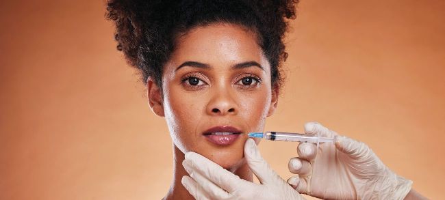
Vascular occlusion (VO) is recognised as the most significant complication associated with soft tissue filler treatments, carrying the potential for permanent tissue loss, scarring, and facial deformity if not promptly identified and managed.9-10 Despite its severity, VO remains relatively uncommon, with reported incidence rates ranging between 0.001% and 0.043%.9-12 This combination of low incidence and high consequence demands that practitioners maintain a comprehensive understanding of relevant anatomy and a high level of vigilance during and after treatment. The risk of VO is influenced by multiple factors, including the filler material used, the injection technique, and the anatomical site involved.9-12
The duration of ischaemia ultimately determines both the medium and long-term outcomes for the patient. Most cases of VO manifest acutely and are therefore identified in-clinic; however, delayed presentations have been documented, sometimes occurring several days after injection.9 In all cases, whether early or late, the Complications in Medical Aesthetic Collaborative (CMAC) recommends attempting to dissolve the filler, even when necrosis and eschar formation have already occurred.9,12 Clinically, the most important examination finding in suspected VO is a capillary refill time exceeding two seconds, which reflects impaired perfusion and should immediately raise concern for arterial compromise.12 CMAC outlines five progressive stages of vascular occlusion, each reflecting escalating tissue injury.13 Early recognition and intervention, ideally before progression beyond the initial stages, significantly improve prognosis and reduce the likelihood of lasting scarring or wound formation.12 Increased pain or disproportionate pain at the injection site can be a potential early onset feature.14-16 Whilst pain isn’t always a feature of VO, its onset – particularly if unexpected – should raise concern. A systematic review assessing filler-induced facial skin ischaemia reported that pain was a more common symptom (77%) than skin discolouration (67%) in 243 cases of vascular occlusion.17
REVIEWING THE LITERATURE:
A structured literature search was carried out using OVID MEDLINE with the search terms dermal filler, hyaluronic acid filler, cosmetic filler, combined with vascular occlusion and skin ischaemia. Only English-language publications were included, and no date limits were applied. The search produced 89 articles. Seven could not be accessed, leaving 82 for review.
Reviewed material comprised a combination of case reports, case series and literature reviews. Of these, 24 papers explicitly discussed the clinical cutaneous signs of vascular occlusion, with some focusing primarily on the clinical presentation of vascular occlusion, whilst others only mentioned it briefly. Notably, only one publication specifically addressed these signs in patients with ethnic skin or skin of colour. The authors of this paper described “the red sign” – punctate dermal bleeding observed immediately after injection – as an indicator of adequate perfusion and absence of vascular compromise.18 The sign was noted to remain dependable even in darker skin tones, where conventional markers (blanching, pallor) are more difficult to assess. They also emphasised the heightened importance of safe injection practices, including the use of microboluses, low pressure, slow injection speed and placement in appropriate anatomical planes to minimise vascular risk in patients with SOC.18
DISCUSSION
Under-representation of SOC
The underrepresentation of SOC in aesthetic medicine extends beyond educational material to include research participation, clinical reporting and professional culture. While SOC patients do seek aesthetic treatments, some may also access procedures through non-medical practitioners, limiting opportunities for data collection, structured follow-up and inclusion in research. Among medical injectors, complication reporting remains low across all patient groups, meaning valuable clinical insights are often lost. In addition, research participation depends on clinicians who both attract and treat diverse populations, undertaking research.
Adverse event reporting
Stigma surrounding adverse events further compounds the problem, discouraging open discussion and shared learning. Creating a culture that normalises the reporting and discussion of complications, without blame or judgement, is essential to improving safety and building an evidence base that represents all skin types. Greater transparency, diversity in research, and inclusive education will collectively strengthen patient care and clinician confidence in aesthetic medicine.
In the UK, adverse events related to dermal filler use should, in principle, be reported to the Medicines and Healthcare products Regulatory Agency (MHRA) via its Yellow Card or medical device vigilance systems. However, available evidence indicates that complications are substantially under-reported. In 2022, the MHRA received only 86 spontaneous reports of adverse incidents associated with dermal fillers, while a broader Freedom of Information (FOI) release identified a total of 1,323 reports across all years to date.9-10
These figures almost certainly underestimate the true incidence, given that reporting is voluntary, depends on individual practitioner initiative, and there is no accurate denominator for the total number of filler procedures performed annually.11
Under-reporting is further compounded by the absence of comprehensive regulation in the UK aesthetic sector. Dermal filler administration is not legally restricted to medical professionals, and practitioners performing non-surgical cosmetic procedures are not required to register with the Care Quality Commission (CQC) unless they also offer surgical interventions.12
As a result, a substantial proportion of procedures are carried out in non-clinical settings, many of which lack robust complication management protocols or formal mechanisms for adverse event reporting. Together, these factors mean that official data almost certainly represent only a small fraction of true complication rates within the UK.
Uptake of cosmetic intervention in patients with SOC
There is substantial and increasing interest in aesthetic procedures from patients with SOC, with the number of surgical and non-surgical cosmetic procedures performed in the US in non-Caucasians increasing from 19% to 25% between 2010 and 2016.8 According to 2020 ASPS statistics, among minimally invasive soft-tissue filler procedures in the US, 78% were performed in Caucasian patients, 10% in Hispanic, 5% in Black or African-American, 5% in Asian or Pacific Islander, and 1% in other ethnic groups.7
Volume loss in SOC
Although patients with SOC, including those of African, Asian and Latino/Hispanic descent, may demonstrate slower or less intrinsic and UV-induced extrinsic facial ageing, these changes still occur and many individuals within these populations seek volume-restorative aesthetic treatments.22-24 In SOC, the most prominent signs of facial ageing are generally observed in the midface and periorbital regions, with comparatively fewer changes in the upper third.23 There is also a reduced tendency towards perioral wrinkles and radial lip lines. Patterns of age-related change vary among ethnic groups. Black patients often present with noticeable sagging of the malar fat pads and early jowl formation.22 East Asian individuals tend to show midface soft-tissue descent, malar fat pad ptosis and tear trough development.22 South Asian patients typically have greater buccal fat and more projected cheekbones, which may confer structural support against midface deflation. Latino populations may exhibit infraorbital hollowing and heavier fat pad accumulation in the midface, causing more prominent nasolabial folds and jowl formation.22 These are generalised observations, and significant individual variation should always be recognised in aesthetic assessment and treatment planning.
Ischaemic signs in patients with SOC
The body of evidence for acute limb ischaemia (ALI) is much more extensive than for filler-related VO, making it a useful comparative framework given the analogous pathology of arterial compromise. ALI is well characterised in terms of clinical presentation. Visual signs of cutaneous hypoperfusion are less obvious in higher-melanin skin.14 Cyanosis may manifest as a grey or violaceous hue rather than classic blue; thus, relying on colour alone is unreliable.14
In darker skin, erythema and livedo patterns are harder to distinguish, reducing the sensitivity of “redness/mottling” as early ischaemic indicators. In one controlled study of induced limb ischaemia, clinicians’ ability to detect ischaemia by visual exam dropped substantially as skin pigmentation increased, suggesting that relying solely on inspection introduces bias.14 Because of these limitations, objective adjuncts should be prioritised across all skin tones. Capillary refill time, temperature asymmetry, Doppler flow signals, and imaging (e.g. ultrasound) offer more reliable assessment of perfusion, especially when visual cues are ambiguous.14
When treating SOC, clinicians should maintain heightened vigilance for any atypical pain response, as both physiological and sociocultural factors can influence how pain is perceived, expressed and interpreted. Evidence shows that pain reported by ethnic minority patients is often underestimated or undertreated, partly due to implicit biases and false beliefs.25-28 Differing cultural norms around the expression and reporting of any pain in certain ethnic groups may further obscure early warning signs of VO. Encouraging cultural awareness, reflective practice and inclusive training is essential to ensure equitable and safe care for all patients.
A recent study explored the use of laser speckle contrast imaging (LSCI), a real-time, non-contact technique that maps tissue blood flow, in the management of filler-induced vascular occlusion.28 In this case series, LSCI accurately identified areas of hypoperfusion following intravascular filler injection and guided targeted needling and hyaluronidase treatment, reducing unnecessary tissue trauma and improving outcomes. The technology allowed for precise, dynamic assessment of perfusion defects, which could offer greater accuracy than visual inspection in patients with SOC. Although LSCI is not yet widely known or available in aesthetic practice, it represents a promising adjunct for diagnosis and monitoring vascular events in real time, which may be particularly valuable when treating patients with skin of colour.
EXPERT OPINIONS
Given the paucity of published information on vascular occlusion, we contacted some expert injectors to seek any anecdotal insights to share to a wider audience.
Mrs Fatou Diouf is a leading aesthetic nurse and founder of the Treat Different educational platform. Based in Ohio, USA, her work focuses on raising standards in aesthetic care for skin of colour through clinically informed training and advocacy. She says ‘The conversation on vascular occlusion often leaves melanin-rich skin out, yet the risks are higher and the signs harder to detect. Our industry must expand its frameworks to protect melanin-rich patients with precision and urgency.’
Mr Ayad Harb is a plastic surgeon based in London, UK. He is an aesthetics educator and has a specialist interest in non-surgical rhinoplasty. He authored the article titled ‘Nonsurgical Rhinoplasty in Patients of African Descent: A Retrospective Review’ quoted earlier in this article. He has treated multiple cases of VO in darker skin and advises that the most reliable sign is the ‘red sign’ described earlier. Relying on discolouration causes false negatives. Treatment protocols for management remain the same, but monitoring recovery is more difficult, so signs of dermal bleeding remain the most reliable signs of adequate perfusion.
Sharon King is an aesthetic nurse and the co-director of ACE (Aesthetic Complications Expert) Group World. She is based in Tamworth, UK. She corroborated the above comments by stating that ‘ashen discolouration seen in ischaemic skin of colour is more likely to go undetected’. We are pleased to hear that ACE Group has added an addendum to its current guideline for managing vascular occlusion, to highlight the key differences for diagnosis in SOC. This is pre-publication and is due to be widely available in early 2026. This addendum will highlight the importance of pain monitoring and CRT in examination. It highlights the necessity for diversity and inclusivity in training and imagery. Finally, it mentions the promising nature of near-infrared spectroscopy (NIRS) as an emerging technology to improve diagnostic accuracy.
CONCLUSION
Vascular occlusion remains a rare but potentially devastating complication of dermal filler treatment. Despite its low incidence, the consequences of delayed recognition and management can be profound, underscoring the importance of practitioner vigilance, anatomical knowledge, and preparedness for immediate intervention. Lessons from other ischaemic pathologies, such as acute limb ischaemia, highlight that in patients with skin of colour, traditional visual indicators of ischaemia are less reliable, necessitating a greater reliance on patient reported symptoms such as severe or progressive pain and measurable signs such as capillary refill time, temperature change, and flow assessment. Awareness of under-representation of SOC inaesthetic medicine resources is improving, and it is encouraging to see that complications groups are making efforts to reflect this by improving diversity and inclusion, and also that emerging technologies provide promise in reducing diagnostic uncertainty in this population. There is a lot of work to do, but we are heading in the right direction.

Scan for references: