Under the surface
Dr Zainab Al–Mukhtar explains why ultrasound scanning has become a vital part of her dermal filler treatment protocol
DR ZAINAB AL MUKHTAR
Dr Zainab Al-Mukhtar is a dental surgeon, advanced aesthetics trainer for Acquisition Aesthetics and owner of Harrow On The Hill Dental & Facial Aesthetics in North London. She is also part of the Beyond Medispa team at Harvey Nichols, Knightsbridge. She qualified at Guy’s King’s & St Thomas’ Dental Institute with Distinction in 2010. Dr Al-Mukhtar was also a facial aesthetics trainer and demonstrator for Oris Medical at the Royal College of General Practitioners. Follow her on Instagram: @drzainabaesthetics
I have been incorporating ultrasound imaging into my clinical practice for hyaluronic acid injectable treatments for around the past three months. My decision to do so was in order to increase safety for my patients. With more people undergoing facial rejuvenation procedures involving volume restoration, facial contouring and augmentation with dermal filler, safety is even more a priority. Luckily, emerging data and equipment in the field makes management of complications more predictable.
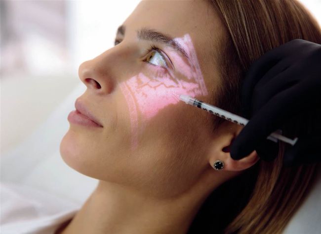
Ultrasound scanning (USS) is a noninvasive way to visualise tissues under the skin. This can be further enhanced by using a colour Doppler scanner to allow for the detection of blood flow. Interest in Doppler ultrasound analysis of the face has grown of late, particularly in the use of injectable fillers. It is now established as a valuable tool in the treatment of filler complications.
One of the rare but serious complications of filler treatments that we aesthetic doctors must be prepared for is a vascular occlusion, in which an artery is obstructed by placement of the filler product, causing restriction of oxygenated blood flow to the surrounding tissues. If left untreated, this leads to ischaemic necrosis (tissue death), the consequences of which can vary from mild scarring to significant disfigurement. Emergency dissolving of filler with hyaluronidase is essential to unblock the obstruction, resuming normal blood flow to the deprived area.
With Doppler USS we can detect the precise location and depth of the problematic filler by assessing where there is a disruption in normal blood flow. This filler can then be targeted under ultrasound guidance and dissolved with hyaluronidase. Without ultrasound, tissues must be flooded with high volumes of hyaluronidase, without knowing the precise location of the blockage. Reversal can still be successful, but cases often need several repeated daily injections of high volumes of hyaluronidase, which can be traumatic for the patient and stressful for the clinician. When unusually complicated vascular occlusions occur, they can be challenging to resolve despite high hyaluronidase volumes, due to the ambiguity of the exact location of the culprit filler bolus.
Having been involved in supporting a few colleagues, who while treating complications used ultrasound imaging, I decided to begin training to use this technology with Dr Leonie Schelke and Dr Peter Velthuis at the Academy of Regenerative Medicine. Under USS guidance, dissolving is conducted with precision and allows for lower hyaluronidase doses, less trauma, and the reassurance of continuous blood flow to the affected area.
TO THE RESCUE
By way of example, a patient was recently referred to me by a colleague. The patient presented with necrosis in her nasal septum after filler injection at a different clinic. Necrosis in the septum is extremely rare, with only one written case report globally so far. 1 This is unlike skin necrosis on the nose which is more widely reported. The unique set of symptoms for septal necrosis include a nosebleed after injection, dental/ palatal pain, crusting inside a nostril or a “stuffy nose”. For rare or complex vascular occlusions like this, ultrasound can be pivotal at locating the exact point of blockage by noting areas of disrupted blood flow and targeting dissolving to reverse the complication.
USS allowed more predictable resolution of the vascular event because through scanning on a colour Doppler, we were able to locate where there was a disruption in normal blood flow in the region and focus dissolving of the hyaluronic acid filler in that location, then re-scan to look for improvements in the blood flow. In the case of septal necrosis this requires tracing the path of flow of the superior labial artery (from the vermillion border of the upper lip up to the columella). Seeing evidence of continuous blood flow was the indication we needed to feel confident that there was no need for more hyaluronidase. The USS supported our assessments of clinical improvement.
Without the USS, it is likely that through dissolving with hyaluronidase and stringent care and follow-ups, the patient would have been eventually resolved of the vascular complication, but this would have taken more time, more dissolving with greater doses and more follow-ups. We would not have been able to trace the flow of blood in the artery and therefore would only be relying on improvement of signs and symptoms.
It is easier to assess improvement in skin necrosis, but in the case of septal necrosis, visualisation and detection of changes inside of the nose is more difficult for injectors and often needs a multidisciplinary approach involving daily ENT (ear, nose and throat) assessments, which in an emergency situation is not always feasible for all injectors. This is why USS is particularly helpful in the management of complex vascular occlusions.
Facial USS can be used for:
• Vascular mapping before or guided injections during filler placement, to prevent intravascular injection
• Optimising aesthetic results
• Monitoring filler changes over time.
NEW SKILLS
Ultrasound imaging is most useful to experienced injectors; a very good understanding of facial anatomy is essential in order to interpret scans successfully.
Training is required to understand ultrasound, and there is a learning curve. Skilful scanning, confidence and interpretation comes with ultrasound experience which takes extensive commitment of time, scanning patients routinely. It is worthy to note that financial, training and time investments are required to use facial USS successfully. For routine use, especially for beginners, additional appointment time is needed. Personally, I now need to add approximately 15 minutes to my filler appointments, especially with nose treatments. It can take anything between two and up to 10 minutes to find the vessels –I have found this to be the case in particularly cold noses. I have also had to increase my prices for non-surgical rhinoplasty in particular to allow for this additional time and attention.
I believe within a few years ultrasound imaging will become standard practice in cosmetic medicine, and time will tell if this comes to pass. However, I for one will be using it as of now as part of the protocol for every filler treatment I undertake.
REFERENCES
1. Souza Felix Bravo, Bruna et al. “Septal Ulcer After Nasal Filling with Hyaluronic Acid.” The Journal of clinical and aesthetic dermatology vol. 14,1 (2021):24-26